Learning points in unit:
The below patient is the first day of my unit duty inthe casuality she came with dengue NS1 positive outside with thrombocytopenia in Tachyponeic state with a petechiae and intact sensorium
Expanded dengue:
It is a terminology developed in the who guidelines 2012
1) unusual manifestations of patients with severe organ involvement such as kidneys,brain ,liver or heart associated with dengue infection have been increasingly reported in dengue hemorrhagic fever or in dengue patients who donot have evidence of plasma leakage. These unusual manifestations may be associated with other co morbidities,co infections, or complications of prolonged shock clubbed under the expanded dengue
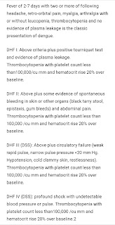
2) Grades of Dengue hemorrhagic fever
2)
Ischemic stroke:
Ischemic stroke is characterized by the sudden loss of blood circulation to an area of the brain, resulting in a corresponding loss of neurologic function.
Acute ischemic stroke is caused by thrombotic or embolic occlusion of a cerebral artery and is more common than hemorrhagic stroke.
Anatomical location in acute ischemic stroke
Middle cerebral artery,
Causes of ischemic stroke
Cardioembolic stroke
Embolus from heart get dislodged in intracranial vessels mca is the most common vessel affected
UMN facial palsy
This picture i have taken from google because icant show the patient face
In central facial palsy, paralysis controlateral to the side of lesion ,eyelid and fore head muscles are not affected
Causes of UMN facial palsy:
Central UMN lesions: eg: stroke
Central facial lesions:
Deviation of uvula to left
The uvula is innervated by the 10th cranial nerve, i.e., the Vagus nerve. The deviation of the uvula to one side may imply two things, in the case of a lower motor lesion of the Vagus nerve on one side, the uvula deviates to the opposite side, but an upper motor lesion of the Vagus nerve on one side causes the uvula to deviate to the same side. For example, a lower motor neuron lesion of the left Vagus nerve causes uvula leaning to the right, but if there is an upper motor lesion of the left Vagus nerve causes the uvula to deviate to the left side itself.
The main causes for the deviation of the uvula are usually due to the weakness in the 9th and 10th cranial nerves.
The infection or compression of these nerves may cause uvular deviation or
it may occur during a stroke when the tongue, the palate and the pharynx are deviated along with the uvula. The compression of the nerves could be caused by a tumor pressing the nerves either outside of the skull or inside the skull, i.e., either before the nerve enters the skull or after it enters the skull.
3)
High out put heart failure
High-output heart failure occurs when the normally functioning heart cannot keep up with an unusually high demand for blood to one or more organs in the body. The heart may be working well otherwise, but it cannot pump out enough blood to keep up with this extra need.
Causes
Thyrotoxicosis
Av fistula,
Chronic Anemia
Pagets disease of bone,beri beri,low systemic vascular resistance
Ejection systolic murmur
Most common kind of heart murmur
Are usually crescendo decrescendo
They may be
Innocent : in children and young adults
Physiologic: can be detected in hyperdynamic states
Eg: Anemia, fever, hyperthyroidism
Pathological: aortic stenosis, pulmonary stenosis
Hypertrophic cardiomyopathy
Iron deficiency anemia causes:
Nutritional,blood loss
Causes of raised jvp:
CardiAc failure
Tr,Ts,cardiac tamponade,constrictive pericarditis
Pulmonary: copd/ cor pulmonale
Abdominal: ascites
4)
Prepyloric gastritis:
Prepyloric local gastritis is a disorder manifested by postprandial epigastric dis- tress and radiologic abnormalities of the distal segment of the stomach
prepyloric local gastritis is primarily a psychosomatic disorder in which the parasympathetic (craniosacral autonomic) nervous system is subjected to excessive stimulation of central origin. The vagus nerves mediate both motor activity and secretion in the stomach. The beneficial effect of vagotomy in some cases of intractable peptic ulcer appears to be established. The administration of atropine, which blocks the nerve endings of the parasympathetic (craniosacral autonomic) system, diminishes or relieves the symptoms of these patients
5)
Diabeticketoacidosis is the most common complication in type 1 diabetes
What are precipitating causes in Dka?
Infection , is most common precipitating cause in known diabetes
In this patient precipitating cause was peri anal abscess and poor glcemic control
How it is diagnosed?
It is diagnosed as a combination of Hyperglycemia, metabolic acidosis, ketonuria
What is the reason for reduced serum sodium levels?
The measured serum sodium levels are reduced as a consequence of osmotic fluid shifts due to Hyperglycemia.( Reduction of 1.6meq for each 5.6mmol/l 100mg/dl rise in serum glucose.
How it was treated?
It was treated by correcting the substantial hypovolemia by giving fluids @ 100ml/hr
Hyperglycemia was treated by giving insulin ( Human Actrapid injection) intravenously
Electrolyte imbalance like hypokalemia is corrected by inj kcl infusion
This picture i have taken from Harrison's principles of medicine
How perianal abscess was treated?
Incision and drainage of abscess under spinal anaesthesia followed by debridement of slough with regular dressings
Conclusion:
Prompt surgical intervention, anti bacterial therapy, rapid restoration of glycemic control are crucial to prevent mortality in diabetes mellitus patient's complicated with abscess
6)
https://60shirisha.blogspot.com/2023/01/liver-abscess.html
Liver abscess
2 main causes:1) amoebic 2) pyogenic
Amoebic :
Organism: entamoeba histolytica
Route. : Flask shaped ulcer- portal vein- liver
Abscess: solitary
Clinical features:
Less toxic , low chance of jaundice
Age: 3rd or 4th decade
Investigations: raised pT,inr
Raised ALP, serology positive for amoeba
IOC: cect
Aspirate: anchovy sauce pus,devoid of neutrophils.
Pyogenic:
Organism: polymicrobial
Route: Ascending cholangitis,or biliary obstruction
Abscess: 50% solitary,50% multiple
Clinical features: more toxic,jaundice is more common,sepsis
Age: 5th decade
Investigations: Raised ALP, raised pt,inr,blood culture positive
IOC: Cect
Aspirate:pus with neutrophils
Management:
Double strength metronidazole 800 mg Tid is started , if the patient is responding it is continued for 2-3 weeks.
A 10 day course diloxanide furoate( luminal amoebocide) is given after metronidazole course
If there is no response:
Aspirate or pig tail catheter insertion
Management of pyogenic liver abscess:
Intravenous antibiotics
If not responding to antibiotics aspiration or insertion of pig tail catheter
Indications of aspiration or pig tail catheter insertion:
Abscess Cavity > 5cm in size
Impeding rupture
Left lobe liver abscess : slim chance of rupture into pericardium
Adequate antibiotic coverage and image guided intervention is the optimal first line treatment with favourable outcome
7)
Why thrombocytopenia is a common in dengue infection
Thrombocytopenia is a common laboratory finding in dengue infection. It usually reaches its nadir during the critical phase and resolves subsequently. The pathophysiology of thrombocytopenia in dengue infection is not clearly understood. It is believed that it rests mainly on two events:
1)decreased in bone marrow production and/or2) increased peripheral destruction and clearance of platelets.
Immune-mediated clearance of antibody-coated platelets has been proposed as one of the mechanisms leading to thrombocytopenia. The cross-reactivity of antibodies directed against NS-1 antigen and platelets suggests the role of antiplatelet antibody in the pathogenesis of thrombocytopenia.In addition, complement-mediated platelets destruction plays an important role during dengue infection.
Why polyserositis is seen in dengue?
Most critical future in dengue remains the leakege of plasma .This leakage of plasma is due to increased endothelial capillary permeability.This may present as ascites,pleural effusion,pedal edema and hemoconcentration.
Conclusion:
The reported women with Dengue fever with thrombocytopenia have been managed by fluids correction based on her hydration status to maintain sufficient urinary output and perfusion
In the above patient poly serositis is due to dengue fever leading to increased endothelial permeability
And plasma leakage
Serositis is the predictor of impending dengue shock syndrome and was treated successfully with fluid replacement therapy.
Once dengue shock syndrome was treated serositis resolves on its own .
8)
Causes of anasarca:
Congestive heart failure
Right failure:
Most common and most serious causes of anasarca
Other causes:
Constrictive pericarditis
Venous obstructions
Increased capillary hydrostatic pressure:
Heart failure
Primary renal disease
Nephrotic syndrome
Acute glomerulonephritis
Renal failure
Decreased capillary oncotic pressure:
Nephrotic syndrome
Liver failure
Protein losing enteropathy
Protein mal nutrition
Increased capillary permeability:
Sepsis
Burns
Other cases in our unit:
A 42 year old female patient
Diagnosis:
1) hypoxic ischemic encephalopathy
2) Generalized status epilepticus secondary to Autoimmune vasculitis- pRES/uremic/ septic encephalopathy
3) pre renal aki on CKD on hemodialysis
4) urinary tract infection
5) Anti synthetase syndrome
6) pulmonary Tb
7)? Invasive aspergillosis
8) post tracheostomy
Clinical pictures of this patient:
Erosion and distorted nails
Hypopigmented patches, blisters
Anti synthetase syndrome:
Anti-synthetase syndrome is an autoimmune disease characterized by autoantibodies against one of many aminoacyl transfer RNA (tRNA) synthetases with clinical features that may include interstitial lung disease (ILD), non-erosive arthritis, myositis, Raynaud’s phenomenon, unexplained fever and/or mechanic’s hands (1). Anti-synthetase syndrome is an idiopathic inflammatory myopathy (IIM), with a higher prevalence of ILD compared to dermatomyositis (DM) and polymyositis (PM), IIMs with which it shares many features. The ILD in anti-synthetase syndrome patients is often severe and rapidly progressive, causing much of the increased morbidity and mortality associated with anti-synthetase syndrome as compared to the other IIMs (2). Diagnostic criteria
| Connors et al. (2010) (1) | Solomon et al (2011) (10) |
|---|
|
|---|
| Required: Presence of an anti-aminoacyl tRNA synthetase antibody | Required: Presence of anti-aminoacyl tRNA synthetase antibody |
PLUS one or more of the following clinical features:Raynaud’s phenomenon Arthritis Interstitial lung disease Fever (not attributable to another cause) Mechanic’s hands (thickened and cracked skin on hands, particularly at fingertips)
| PLUS two major or one major and two minor criteria:
Major:Interstitial Lung Disease (not attributable to another cause) Polymyositis or dermatomyositis by Bohan and Peter critieria
Minor:Arthritis Raynaud’s phenomenon Mechanic’s hands
|
PRES ( posterior reversible encephalopathy syndrome)
r A rarecondition marked by headaches, vision problems, mental changes, seizures, and swelling in the brain. The symptoms of PRES usually come on quickly and can be serious and life threatening. When treated, symptoms often go away within days or weeks. PRES may occur in patients with certain conditions, such as high blood pressure, eclampsia, severe infection, kidney disease, and certain autoimmune diseases. It may also occur in patients treated with certain anticancer drugs and immunosuppressive drugs. Also called posterior reversible encephalopathy syndrome, reversible posterior leukoencephalopathy syndrome, and RPLS.
Differences between locked in syndrome and vegetative state
The difference between locked-in syndrome and a vegetative state is that a person with locked-in syndrome retains their full mental faculties, whereas a person in a vegetative state does not. However, because locked-in syndrome causes the loss of all physical capabilities and all muscle movement other than the eyes, people can easily mistake it for a vegetative state
✓85year old female came with complaints of pain in abdomen
C/o burning sensation in chest since 2months
And lower back pain since 2months
C/o loss of appetite since 5-6months
Patient was apparently alright 5-6months ago then she started experiencing loss of appetite
Patient started experiencing burning in the chest since 2months
Is associated belching and burning sensation in abdomen
And occasional pain in right iliac region, relieves spontaneously and lower back pain since
Burning sensation aggravates on eating tangy food or dal, early satiety since 5-6months
Burning sensation relieves for a while on having food
No radiation of pain, shortness of breath , palpitations and profuse sweating
No h/o nausea, vomiting, weight loss
K/c/o hypertension since 1 year (patient don’t the medication name)
In this in USG finding there is a E/O multiple small hyperechoic foci in both lobes of liver
DD'S: calcification s / granuloma
2) von meyenburg complexes
Von Meyenburg Complexes (VMCs) is a rare clinicopathologic entity, consisting of small (<1.5cm), usually multiple and nodular, cystic lesions, which are caused from ductal plate malformations of the smallest intrahepatic bile ducts. These lesions typically cause no symptoms or disturbances in liver function and thus, in most instances, VMCs are diagnosed incidentally, on the basis of their unique radiologic appearance.
learning points in ICU:
First day of my ICU night duty:
I have seen the patient who was on mechanical ventilator who is diagnosed with sepsis with mods and right heart failure . His bp was not recordable in the morning and pulse was feeble and he was sedated and ionotropes were given .
In between he had fever spikes .
Another patient : 26 year old male with
CKD ON MHD since 3 months
Left lung empyema ICD- 10
Uncontrolled hypertension
K/c/o hypertension since 3 months
S/P:RT nephrectomy 10 years ago
Hypertensive retinopathy
This patient had high bp recordings like 220/160 mmHg evening, early morning time
2 nd day : i have seen altered sensorium case
was altered sensorium secondary to hypotonic hyponatremia? Siadh
3rd day: a patient on 3rd day ventilator sedation has with hold and he had spontaneous breathsof 40 - 45 and his heart rate was increasing around 130-140 bpm and the secretions were drained and in mid patient pulses were not palpale cpr was initiated for 20 minutes and Rosc was obtained and pulse was disappeared again ionotropes ,atropine were given his rate was increased but pulses were not palpale cpr continued and the patient was died .
Immediate cause - VF ( ventricular fibrillation).
Antecedent cause - Post CPR status
Acute decompensated heart failure with reduced ejection fraction ( EF - 30% )
? Old CAD
S/P papillary thyroid carcinoma
Acute kidney injury
4th day:
One patient came wth severe sob with lower limb cellulits
And his abg was done which showed severe metabolic acidosis and bicarbonte 20 meq/l given over 20 minutes

And his spo2 was 96% under 6 litres of oxygen
And in the rounds i have seen the patient in imaginary pillow .
This patient underwent many Hemodialysis sessions and 2 times intubated and extubation has done and again re intubated due to low saturation levels
Procedure : Ascitic tap was done in z track technique under guidance of Dr Haripriya mam
Last day : i have monitered one patient whose bp were not recordable and pulses were feeble and started on ionotropes and sedation was given
And in evening her pulse rate was increased to 160 BPM : ( ventricular tachycardia)
Diltiazem was given and her heart rate decreased to 123bpm.
Below is the case:
This is a case of 40 year old female, who presented to casuality with poor GCS ( E1V1M1) in Gasping state, with Bp,PR not recordable, and SpO2 60% on monitor, with absent central pulses. Immediate cpr was initiated and patient was intubated meanwhile, and after 25 min of CPR ,ROSCattained and inview of not recordable Bp , patient was started on inotropes, and pupils were dilated and fixed, not reacting to light and MRI brain was done, after attaing of BP : 80 mmHg which showed subarachnoid hemorrhage and intraventricular hemorrhage with raised ICP and mild tonsillar herniation. Neurosurgery opinion was taken and advised to continue conservative management .echo showed LAD ,RCA+ LX hypokinesia with mild to moderate LV dysfunction. Patient had refractory hypotension, inspite. Of multiple ionotropic support and brain stem reflexes were absent
On 27/12/22 ,6.15 AM patient had sudden cardiac arrest , Rosc attained at 6:35 AM and at 7:00 AM, patient had sudden cardiac arrest , cpr continued for 45 min
Inspite of all resuscitative efforts , patient could not be revived and declared dead at 7:48 Am on 27/12/22
Antecedent cause : Diffuse sub arachinoid hemorrhage with intraventricular hemorrhage with mild acute hydrocephalus with mild tonsillar herniation with raised intracranial tension with post cpr status ( 2times) - Rosc 25 mins - 1st episode
Rosc 2 nd episode - 20 min with heart failure with reduced ejection fraction secondary to ischemia with cardiogenic shock with dural venous sinus thrombosis ??
Immediate cause:
Cardiogenic shock
Tonsillar herniation
Along this case ihave learned some basic things
Vitals monitering
Setting up iv drip set
Iv injections
Passing rules tube
Foley's catheter insertion
Abg sample collection and analysis
Ventilator settings
Emergency drugs:
Adrenaline, atropine
Lumbar puncture procedure
Nebulization
Cpap,bipap,
Endotracheal intubation
Intubation and extubation criteria
Indications
Cpr
Icu check list:
Feeding
Analgesia
Sedation
Thromboprophylaxis
Head of bed elevation
Ulcer prophylaxis
Glucose control
Spontaneous breathing trial
Bowel care
Indwelling catheter removal
De Escalation of antibiotics
Nephrology learning points:
On first day i have seen how to do centralline via femoral artery and assisted Dr.chandana mam and learned how to do procedure and instruments

On night a patient of 46 year old male known case CKD on MHD with irregular Hemodialysis sessions was complaining of unilateral hip pain and burning sensation in stomach and abdominal pain. He had recurrent hypoglycemic episodes 25 d was connected and in morning patient was shifted to dialysis on that time his saturation levels are below 70 and pulses was not palpable and bp was not recordable and the patient was shifted to icu and the patient was sedated and intubated . And his bp recordings were only heared only during systolic around 100or90mmhg and at night patient was shifted to dailysis upto 30 minutes his vitals were normal and after that his body had become cold and colour changes of body, stiffness of hands and sweating, breath rate 60 cpm, tachycardia 140 BPM, and the pulses were not palpable cpr was initiated for around 20 minutes and automated external defibrillator was initiated and patient not responded and declared as death
Renal biopsy
I have learned how to do renal biopsy procedure under USG guidance
Dialysis
Ward :
20 year old female came to the opd on 17/12/22 with complaints of generalised swelling of body since 45 days
Hopi : The patient was apparently asymptomatic 5 months back then she developed pruritic lesions all over the body and healed with hyperpigmented lesions and in different stages of healing .The patient then developed generalised swelling of body starting initially with bilateral pedal edema which was pitting type insidious onset and gradually progressive. To abdomen
C/o facial puffiness
H/O decreased urine output, then patient went to local hospital and was treated .
Patient now presented with b/l pedal edema upto knees which was pitting type
No h/0 sob, cough, ,vomiting, loose stools
General examination:
Patient was conscious, coherent cooperative
Pallor: present
Renal biopsy was done under strict aseptic conditions on 21/12/2022
SPOT URINARY PROTEIN: 5.45
SPOT
SPOT URINARY PROTEIN: 5.45SPOT U2RINARY CREATININE: 21
RATIO: 0.25
24
24 HR URINARY PROTEIN- 436 mg/day
24 HR URINARY CREATININE- 0.8 gm /day
24 HR URINE VOLUME- 800 ml
CREATININE: 21
RATIO: 0.25
24 Diagnosis: Nephrotic syndrome under evaluation with anemia of chronic inflammation and Denovo hypertension and hyper trophic lichen planus:
SPOT URINARY CREATININE: 21
24 HR
Diabeticketoacidosis is the most common complication in type 1 diabetes
What are precipitating causes in Dka?
Infection , is most common precipitating cause in known diabetes
In this patient precipitating cause was infection, myocardial infarction ( old inferior wall)
How it is diagnosed?
It is diagnosed as a combination of Hyperglycemia, metabolic acidosis, ketonuria
What is the reason for reduced serum sodium levels?
The measured serum sodium levels are reduced as a consequence of osmotic fluid shifts due to Hyperglycemia.( Reduction of 1.6meq for each 5.6mmol/l 100mg/dl rise in serum glucose.
How it was treated?
It was treated by correcting the substantial hypovolemia by giving fluids @ 100ml/hr
Hyperglycemia was treated by giving insulin ( Human Actrapid injection) intravenously
Electrolyte imbalance like hypokalemia is corrected by inj kcl infusion


Comments
Post a Comment